The Latest in Live Cell Imaging Biology Diagrams
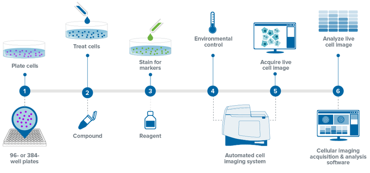
The Latest in Live Cell Imaging Biology Diagrams 1.2 Live-Cell Imaging Considerations 1.2.1 Environment. Maintaining cell health is critical in live-imaging experiments. Mammalian cells must be viable and healthy during the imaging process to ensure that they will progress normally through the cell cycle. Important parameters that must be maintained include temperature, osmotic pressure and pH.
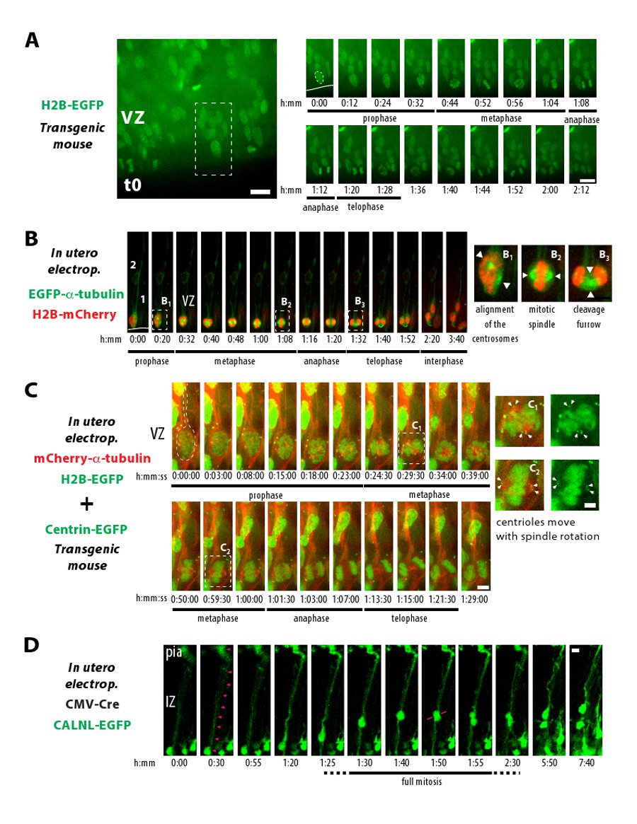
Live cell time-lapse imaging is an important tool in cell biology that provides insight into cellular processes that might otherwise be overlooked, misunderstood, or misinterpreted by the fixed-cell analysis. including the identification of cells at various stages in mitosis, identification and tracking of mitotic defects, and analysis of
Mitosis Through the Microscope: Advances in Seeing Inside Live ... Biology Diagrams
For live imaging, use a temporal resolution of less than 5 min to enable the identification of the different phases of mitosis. Using Syto11 and/or histone H2B-EGFP, 4 - 5 hr of live imaging are sufficient to observe a significant number of mitotic cells.
Mitosis is a highly dynamic and choreographed process in which chromosomes are captured by the mitotic spindle and physically segregated into the two daughter cells to ensure faithful transmission of the genetic material. Live-cell fluorescence microscopy enables these dynamics to be analyzed over diverse temporal scales.
Imaging mitotic processes in three dimensions with lattice ... Biology Diagrams
By comparing mitosis in live wild-type and mutant cells, it has been shown, e.g., that cytoplasmic dynein is required for positioning the spindle in yeast , for the information lost in the undersampled data sets acquired during live cell imaging. This means that such methods can improve the resolution of the original fluorescence image and 73.1K Views. Mitosis is a form of cell division in which a cell's genetic material is divided equally between two daughter cells. Mitosis can be broken down into six phases, during each of which the cell's components, such as its chromosomes, show visually distinct characteristics. Advances in fluorescence live cell imaging have allowed scientists to study this process in great detail In this long history of cell biology of mitosis, however, whole-cell three-dimensional (3D) live imaging with sufficient spatiotemporal resolution to discriminate nanoscale-sized mitotic machineries and completely track their movements has never been achieved. Liu Z, Lavis LD, Betzig E (2015) Imaging live-cell dynamics and structure at the